AsianScientist (Oct. 16, 2015) – For the first time, researchers have recreated two key processes essential for fetal formation in vitro. The team from the Agency for Science, Technology and Research’s (A*STAR) Institute of Bioengineering and Nanotechnology (IBN) has not only controlled the differentiation of stem cells into other cell types, but they have also demonstrated the successful migration of these transformed cells. Their results, published in Scientific Reports, makes it possible for researchers to build a better embryo development model for testing drugs causing birth defects.
Teratogens are compounds that are known to cause malformation in embryos. Currently, researchers rely on animal testing to assess the hazard of teratogens, but this method is expensive, time-consuming and unreliable due to inter-species variability. To overcome these problems, researchers have focused on developing alternative tests using human pluripotent stem cells (hPSCs).
According to IBN Group Leader Professor Hanry Yu, “Embryonic development does not only consist of the transformation of stem cells into other cell types, such as bone, muscle or nerve cells. It also involves the migration of these transformed cells to the right places in the body where they will develop into properly functioning organs as intended.”
“This is why we believed it was important to develop a model encompassing both cell differentiation and migration that would give us a more complete and accurate picture of the effects of teratogens on the developing embryo.”
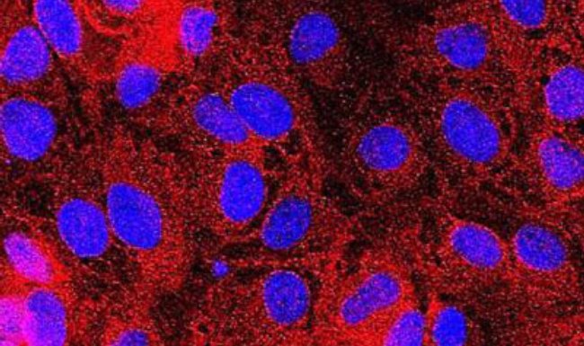
The researchers confined the environment in which the embryonic stem cells transformed into other cell types, and restricted the ensuing migration of the micropatterned hPSC colonies so that the resulting mesoendoderm cells would form a consistent circular or ring pattern. Due to this geometric restriction, it was possible for the researchers to study the effect of teratogens, which may alter the shape and even the eventual position of the mesoendoderm layer.
“A key feature of our model that would facilitate its application as a drug screening platform is the consistency with which we can generate the circular mesoendoderm layer. This provides a reliable starting point and straightforward indicator for measuring drug-induced effects,” said Yu.
The researchers also developed image processing and statistical algorithms to quantify and classify the teratogenic potential of different compounds. Using their micropatterned hPSC model, they have successfully distinguished between teratogenic compounds (i.e. Thalidomide) and non-teratogenic compounds (i.e. Penicillin G), and could also measure dose-dependent effects, which is essential for identifying a teratogenic agent’s clinically relevant dose.
The article can be found at: Xing et al. (2015) A Method for Human Teratogen Detection by Geometrically Confined Cell Differentiation and Migration.
———
Source: A*STAR.
Disclaimer: This article does not necessarily reflect the views of AsianScientist or its staff.