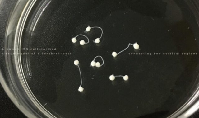
AsianScientist (Nov. 7, 2017) – In a study published in Stem Cell Reports, scientists have invented a microdevice that enables the formation of bundles of nerve fibers, known as fascicles, in the lab.
Axons, or nerve fibers, are the structures through which neurons transmit information to other cells. In the body, they aggregate to form fascicles. Several technologies allow scientists to generate and study single axons in the lab, but none are effective at creating nerve fascicles.
Moreover, although many scientists have examined axon development and degeneration in two-dimensional (2D) systems, it is becoming increasingly apparent that the fascicle’s 3D structure has an essential role in axon function. Fascicles are disrupted in many neurodegenerative diseases such as amyotrophic lateral sclerosis; hence, the understanding of fascicle formation could give clues on the prevention of a number of diseases.
In this study, a collaboration between researchers in Japan and the United States has resulted in the creation of a microdevice that allows the successful formation of fascicles in the lab.
“We know that growing axons form fascicles, but we do not know how fascicles form,” said Dr. Yoshiho Ikeuchi, a lecturer at the Institute of Industrial Science at the University of Tokyo and senior author of the study.
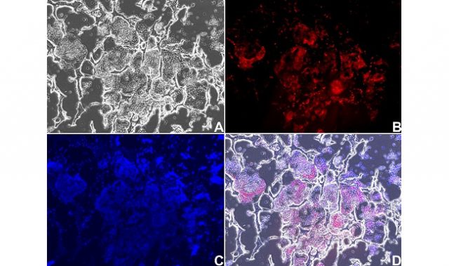
To form axon fascicles, the research teams manufactured a microdevice in which human neurons derived from induced pluripotent stem cells were injected. The stem cells formed neural spheroids inside the chamber of the device.
Axons grew from these spheroids, with some entering the microchannel. Upon this entry, other axons would spontaneously follow, leading to the formation of fascicles that showed properties consistent with those seen in brains. The molecular signaling that caused the spontaneous entry remains unexplained, but fascicles were detected in more than 90 percent of experiments.
“The device gives us a means to investigate which factors are responsible for the fascicle assembly,” said Ikeuchi.
Accordingly, they simulated neurodegenerative conditions by introducing peroxide into the channels, and the fascicles responded with morphological changes representative of the disease state.
These findings and the relative ease of the experiments suggest that the microdevice can be used to test experimental drug compounds that prevent fascicle degeneration caused by disease.
The article can be found at: Kawada et al. (2017) Generation of a Motor Nerve Organoid with Human Stem Cell-Derived Neurons.
———
Source: University of Tokyo.
Disclaimer: This article does not necessarily reflect the views of AsianScientist or its staff.