AsianScientist (Jan. 2, 2018) – In a study published in the journal Proceedings of the National Academy of Sciences, a research group in China has created a three-dimensional (3D) map of neurons in the brain involved in regulating the senses, movement and behavior.
The cholinergic system in the brain plays crucial roles in regulating sensory and motor functions, as well as cognitive behaviors, by modulating neuronal activity throughout the cortex and subcortical nuclei. Dysfunction of cholinergic neurons leads to brain diseases such as Alzheimer’s disease and sleeping disorders. The generation of a whole-brain atlas for the cholinergic system is essential for understanding the functions of cholinergic neurons.
However, previous studies based on 2D sectioning and immunostaining only provided the rough distributions and estimated number of cholinergic neurons. Because of the lack of precise annotation and accurate identification of cholinergic neurons with their branching patterns, the manner in which cholinergic neurons innervate different brain regions is still obscure.
In the present study, a team of scientists led by Dr. Luo Qingming and Dr. Gong Hui’s team at Huazhong University of Science and Technology, and Dr. Qiu Zilong’s Lab at the Institute of Neuroscience of the Chinese Academy of Sciences, provided a precise 3D whole-brain atlas for the cholinergic system and the complete morphology of individual cholinergic neurons in the basal forebrain. Their findings provide a valuable resource for classifying neuronal cell types based on their genetic labeling, connectivity and morphology.
To generate the precise anatomical information of individual neurons, the researchers took advantage of transgenic mice and a virus labelling technique to specifically label the cholinergic neurons in mouse brain. They then applied their whole-brain imaging system, known as the brain-wide positioning system (BPS), to reveal stunning neuronal distribution and morphologies of several representative populations of cholinergic neurons. Their method allowed them to obtain even information about the precise location, numbers, density and soma volumes of cholinergic neurons in specific brain areas.
The morphology of 50 cholinergic neurons in the basal forebrain, reconstructed with the high-resolution imaging of dendritic and axonal processes, showed that adjacent cholinergic neurons can project to multiple distinct nuclei, which contradicts previous hypotheses. The researchers thus suggested a new organized model of cholinergic projections, in which individual cholinergic neurons tend to project to interconnected brain areas.
Looking forward, this 3D atlas may shed light on brain disorders associated with the brain’s cholinergic system and spark inspiration for more detailed cholinergic neuron classifications.
The article can be found at: Li et al. (2017) Generation of a Whole-brain Stlas for the Cholinergic System and Mesoscopic Projectome Analysis of Basal Forebrain Cholinergic Neurons.
———
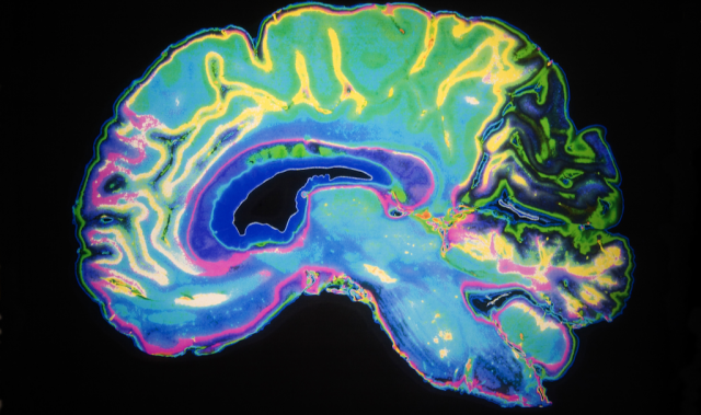
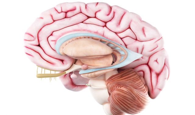
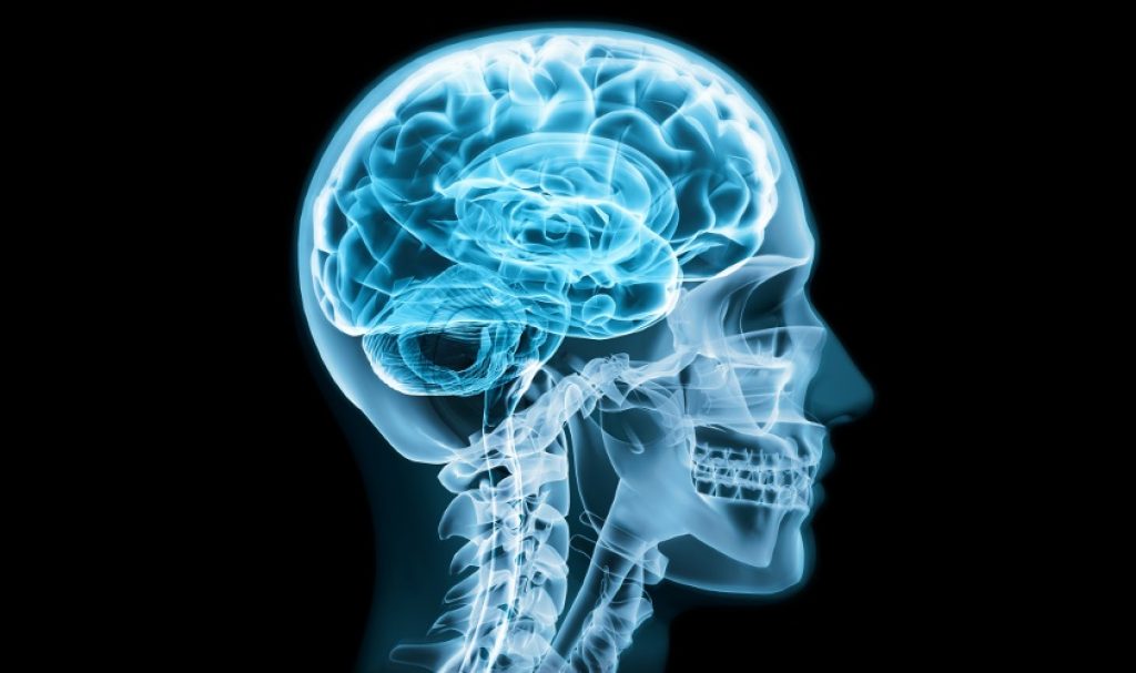
Source: Chinese Academy of Sciences; Photo: Shutterstock.
Disclaimer: This article does not necessarily reflect the views of AsianScientist or its staff.