AsianScientist (May 6, 2015) – Researchers at Kobe University have developed a safe surgical clip that dissolves and be absorbed by the body after a certain period of time. Clinical use of this clip is expected to reduce the rate of postoperative complications and minimize problems associated with diagnostic imaging.
The clip was developed as a collaboration between the Division of Mechanics and Physics of Materials at the Kobe University Graduate School of Engineering and the Division of Hepato-Biliary-Pancreatic Surgery at the Kobe University Graduate School of Medicine.
Most surgical clips are currently made of titanium and as many as 30 to 40 clips may be used during a single surgical procedure. They remain inside the patient’s body after the wounds are healed. Retained clips lead to diminished quality of CT (computed tomography) and MRI (magnetic resonance imaging) images around the wound and may cause complications.
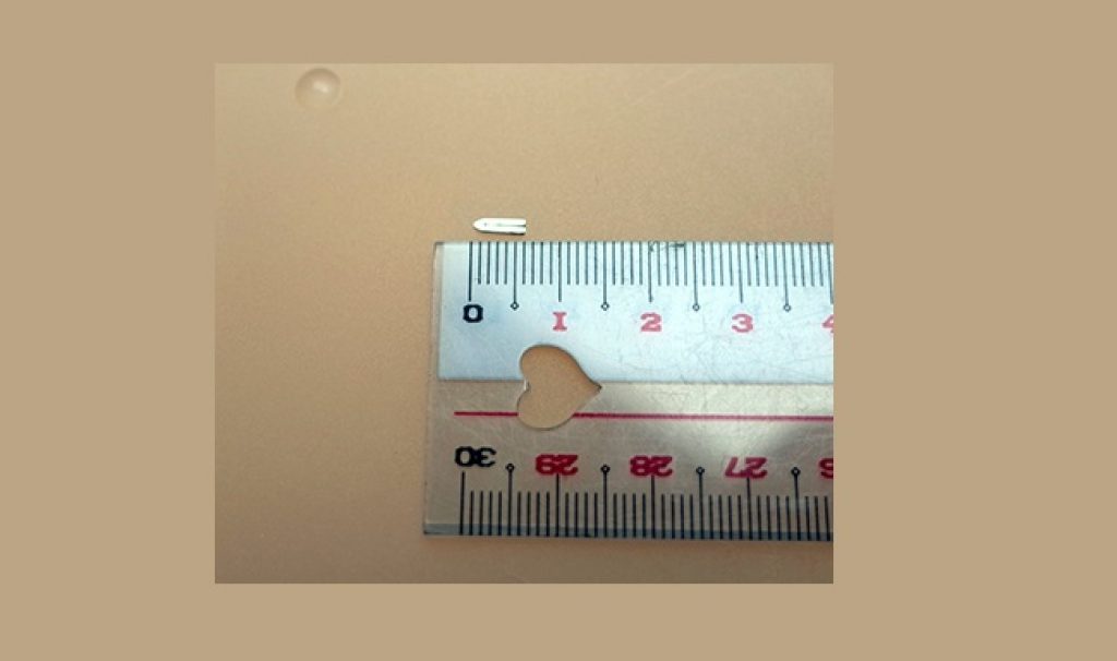
The newly developed clip is five millimeters in size and made of a magnesium alloy. The alloy also contains calcium and zinc to improve its microstructure, ensuring fastening ability and formability, qualities required of materials to make clips.
The safety and functionality of the clip were evaluated in vivo studies. To evaluate the safety, an implantation study was conducted in a subcutaneous mouse model. Very little gas was produced as the clip dissolved and there was no inflammation of the surrounding tissues after one to 12 weeks.
Blood testing revealed that levels of magnesium and other substances in the blood were in the normal range after 12 weeks. The volume of the implanted clip was reduced by almost half after 12 weeks. Therefore, the clip is likely to dissolve and exit the body within one year.
The clip was also tested in a rat model in which the biliary duct, portal vein, hepatic artery and hepatic vein were occluded with the clip and a partial liver was removed. The rat had no problems during a monitoring period of eight weeks, suggesting that the clip functioned properly. Micro CT scanning of the mouse and rat revealed that the quality of images was not degraded and organs can be observed.
“We will conduct further in vivo studies and a clinical study within two to three years,” said Professor Toshiji Mukai, who was involved in developing the clip.
———
Source: Kobe University.
Disclaimer: This article does not necessarily reflect the views of AsianScientist or its staff.