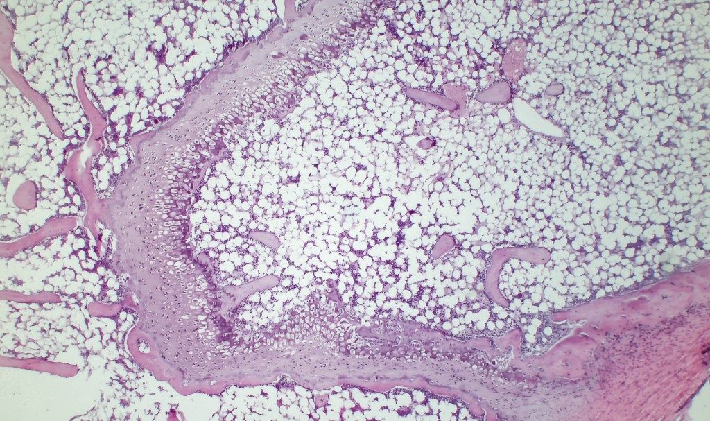
AsianScientist (Jun. 09, 2026) – Bone marrow is found inside nearly every major bone in the body, producing billions of blood cells, platelets and immune cells each day. But before marrow can form during foetal development, the body must carve out space within the developing skeleton. Long bones begin as solid structures, and parts of that dense interior must be cleared to make room. Without this hollowing process, marrow cannot take hold.
The cells responsible are osteoclasts, which break down cartilage and bone to widen the growing cavity. Their activity depends on a signalling protein called RANKL. Without it, osteoclasts fail to develop, and the marrow cavity never forms properly. Although RANKL’s role in bone marrow formation was already known, which cells produce it during foetal development and in what sequence remained unclear.
A study from Kyushu University in Japan now suggests that bone marrow formation unfolds through a developmental handoff between two specialised cell populations. One group initiates the hollowing process inside the developing bone, while another expands and maintains the growing marrow cavity. This two-step mechanism is detailed in the study published in Nature Communications.
To identify the cells, the team engineered reporter mice that allowed RANKL-producing cells to be tracked throughout bone development. They combined single-cell RNA sequencing, which reads the genetic activity of individual cells, with genetic lineage tracking to determine which cells produced the protein, when they emerged and how their roles changed over time.
The earliest RANKL-producing cells emerged from the perichondrium, the connective tissue surrounding the cartilage, and migrated inward along invading blood vessels towards the boundary between cartilage and newly forming bone.
Single-cell sequencing identified these as CD55⁺CD90⁺ mesenchymal cells, a previously uncharacterised population of connective tissue cells originating from the perichondrium. Their genetic profile closely resembled that of septoclasts, specialised cells that secrete enzymes to dissolve cartilage matrix. The study found that these early cells drive osteoclast formation and later give rise to septoclasts during perinatal development, helping establish the conditions needed for marrow formation. When the team deleted RANKL specifically from the population, osteoclast numbers fell sharply, and cavity expansion was delayed.
“Just as lymphoid tissue inducer cells initiate lymph node formation, we hypothesised that a distinct cell population may similarly serve as an organiser to trigger the formation of the bone marrow cavity,” said Shinichiro Sawa, a Professor at Kyushu University’s Medical Institute of Bioregulation.
As foetal development progressed, the early population receded. Leptin receptor-positive bone marrow stromal cells (LepR+ BMSCs) took over as the primary source of RANKL, sustaining osteoclast activity through late foetal development, adolescence and into adulthood. Fate-mapping experiments showed that around 80% of these stromal cells in three-week-old mice originated from progenitors that emerged after the first wave, confirming that the two populations act in sequence.
The study also found that the same sequence reappears during fracture repair. Following injury to the tibia, septoclast-like cells and marrow stromal cells accumulated again near healing tissue and produced the same osteoclast-supporting signals observed during foetal development. The results suggest that fracture healing reactivates a cellular programme first used to build the marrow cavity in the embryo.
The findings have direct implications for bone diseases driven by excessive osteoclast activity, including osteoporosis and rheumatoid arthritis. Current treatments mainly target osteoclasts or RANKL directly, and targeting the upstream stromal cells that regulate osteoclast activity could offer an earlier point of intervention in the processes that drive bone loss.
—
Source: Kyushu University ;Image: BioFoto/Shutterstock
This article can be found at: Medullary cavity expansion is mediated by distinct cell populations during fetal bone development
Disclaimer: This article does not necessarily reflect the views of AsianScientist or its staff.