AsianScientist (May 15, 2019) – Researchers in South Korea and Singapore have developed a chemical probe that enables imaging of a type of immune cells in the brain, known as microglia, in a living animal. The research is published in Angewandte Chemie.
Microglia are the brain’s primary resident immune cells. Although microglia were described a century ago, these cells have only recently been found to play an important role in the development of various neurological diseases.
Discoveries about the link between microglia and neurological disorders have been aided by technological advances in isolating microglia, in conjunction with transgenic small animal systems that express fluorescent proteins from microglia lineages, allowing for live imaging with light microscopy. However, no current methods allow the visualization of microglia at cellular resolution in a live brain, which is clinically more relevant.
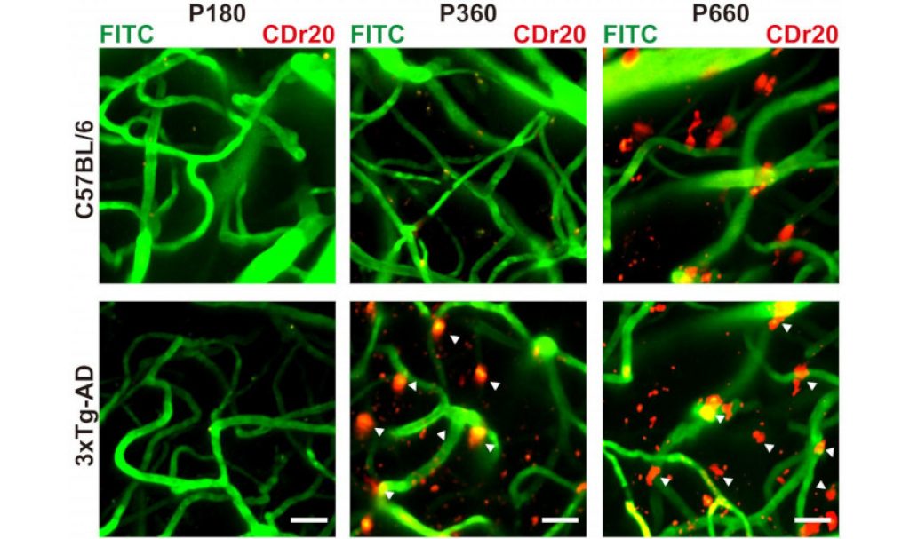
In this study, scientists led by Professor Chang Young-Tae at the Institute for Basic Science, South Korea, with colleagues in Singapore, identified a fluorescent marker for microglial cells, screened from a library of potential probes. The researchers also demonstrated that one of the probes, named CDr20, could indeed label microglia in live brain. Immunostaining was used to verify the selective staining of microglia.
The authors then used knock-out cell lines to discover the enzyme present in microglia that ‘switched on’ the fluorescence of the probe.
“Using a genome-scale CRISPR-Cas9 knockout screen, we identified Ugt1a7cas the functional target protein of CDr20 that activates the CDr20 fluorescence signal in microglia through the enzymatic glucuronidation reaction. Our probe can also label human and primate derived microglia, so this will be extremely useful to study microglia function in higher mammals, which is clinically more relevant.”
The authors noted that they will continue to improve the functionality and utility of CDr20. Furthermore, the Chang laboratory is screening new molecules that only label activated microglia, which scientists suspect to play a role in neuroinflammation in neurodegenerative disorders.
The article can be found at: Kim et al. (2019) Visualizing Microglia with a Fluorescence Turn‐On Ugt1a7c Substrate.
———
Source: Duke-NUS Medical School; Photo: Masahiro Fukuda/Duke-NUS Intravital Imaging Core.
Disclaimer: This article does not necessarily reflect the views of AsianScientist or its staff.