AsianScientist (Oct. 29, 2018) – Researchers at the Okinawa Institute of Science and Technology (OIST), Japan, have imaged the structure of a key component of the Ebola virus at near-atomic resolution. They reported their findings in the journal Nature.
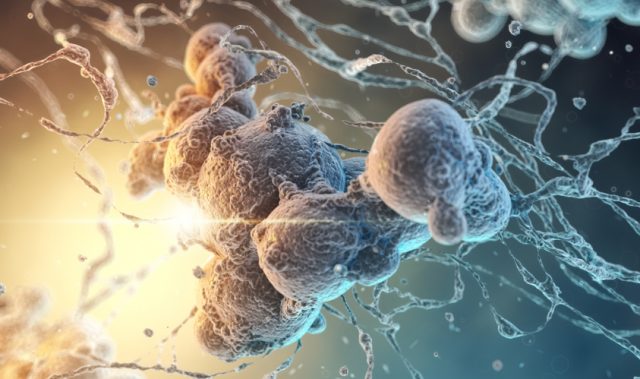
Ebola is a severe disease characterized by symptoms such as fever, fatigue, diarrhea and vomiting, and can lead to death if left untreated. While it has been largely eliminated in the developed world, a recent resurgence of the Ebola virus has occurred in Africa. The virus is transmitted to humans by direct contact with blood or other bodily secretions of infected animals, such as fruit bats and chimpanzees, after which human-to-human transmission is possible.
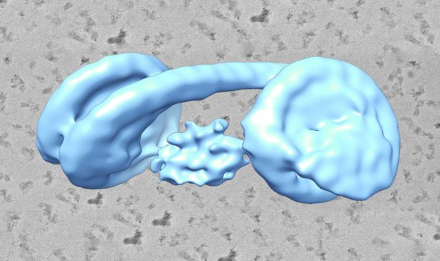
In the present study, researchers led by Professor Matthias Wolf and Dr. Yukihiko Sugita of OIST’s Molecular Cryo-Electron Microscopy Unit (MCEMU) have obtained an ultra-high-resolution image of the Ebola virus’ structure. They focused on a part of the virus called the nucleocapsid (NC), a protein complex that acts as a support structure for the viral genetic material. This structure allows the virus to replicate its deadly payload.
Previous studies analyzed the NC structure using a technique called electron tomography, but the cryo-EM reconstruction generated by the OIST team has much higher definition, resolving individual RNA nucleotides and amino acid side chains.
The researchers first isolated nucleoprotein-RNA complexes, which make up the core of NCs, then analyzed them with a cryo-electron microscope (cryo-EM). They then took more than 18 months to find the right way to reconstruct the NC as a digital model, eventually piecing together the three-dimensional helical structure of the protein complex.
“Before this study, we only knew about the smallest pieces of the NC structure,” said Sugita, “Now that we can see it as a whole, it may help find targets for antiviral drugs.”
The researchers highlighted that neither they nor the public were in any danger from the study. The nucleoprotein-RNA complexes were produced without any actual viral RNA sequence, in non-infectious cells grown in the laboratory at OIST under proper biosafety conditions.
“It’s like the parts of a car,” said Wolf. “Unless the chassis, engine, wheels and so on are all together, nothing works. That’s what the particle we studied is like—most of its components are absent. It’s harmless.”
The three-dimensional model generated from the cryo-EM images provides an accurate foundation for future work. Researchers will now be able to make precisely targeted studies of the entire structure of Ebola virus NC, which may reveal ways to defeat the virus.
The article can be found at: Sugita et al. (2018) Cryo-EM Structure of the Ebola Virus Nucleoprotein–RNA Complex at 3.6 Å Resolution.
———
Source: Okinawa Institute of Science and Technology Graduate University.
Disclaimer: This article does not necessarily reflect the views of AsianScientist or its staff.