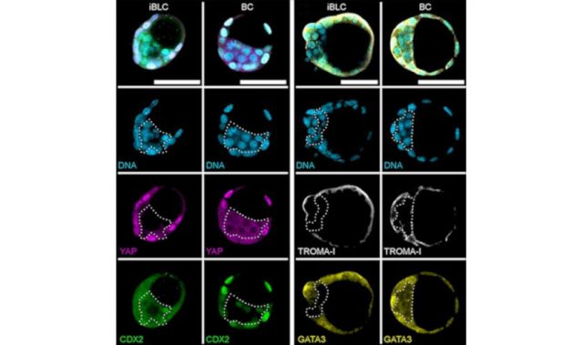
AsianScientist (Apr. 20, 2017) – Stem cell differentiation can now be seen thanks to a combination of machine learning and microfabrication techniques developed by scientists at the RIKEN Quantitative Biology Center in Japan. The results, published in PLOS ONE, followed the differentiation of human mesenchymal stem cells (MSC) which are easily obtained from adult bone marrow.
MSCs have proven to be important for regenerative medicine and stem cell therapy because they can potentially repair many different types of organ damage. Depending on the way the cells are grown, the results can be quite different, making controlling differentiation is an important goal.
Observing MSC differentiation under different conditions is an essential step in understanding how to control the process. However, this has proved challenging on two fronts. First, the physical space in which the cells are grown has a dramatic impact on the results, causing significant variation in the types of cells into which they differentiate. Studying this effect requires consistent and long lasting spatial confinement. Second, classifying the cell types which have developed through manual observation is time consuming.
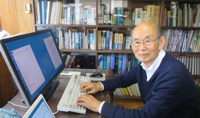
Previous studies have confined cell growth with fibronectin on a glass slide. The cells can only adhere and differentiate where the fibronectin is present and are thus chemically confined. However, this procedure requires high technical skill to maintain the confinement for an extended period of time. To overcome this, the first author of the study, Dr. Nobuyuki Tanaka, decided to look for a new way to confine them. Using a simple agarose gel physical confinement system, he found that he could maintain them for up to 15 days.
“It was wonderful to be able to do this, because agarose gel is a commonly used material in biology laboratories and can be easily formed into a micro-cast in a PDMS silicone mold,” Tanaka said.
“The advantage of this system is that once the PDMS molds are obtained the user only needs agarose gel and a vacuum desiccator to create highly reproducible micro-casts.”
Tanaka’s paper also describes an automated cell type classification system, using machine learning, which reduces the time and labor needed to analyze cells.
“Combined together, these tools give us a powerful way to understand how stem cells differentiate in given conditions,” he added.
The article can be found at: Tanaka et al. (2017) Simple Agarose Micro-confinement Array and Machine-learning-based Classification for Analyzing the Patterned Differentiation of Mesenchymal Stem Cells.
———
Source: RIKEN; Photo: Shutterstock.
Disclaimer: This article does not necessarily reflect the views of AsianScientist or its staff.