AsianScientist (Jun. 1, 2016) – A team of scientists in Singapore has developed advanced microscopy techniques to monitor embryo development in real time, revealing how mammalian cells differentiate during the earliest stages of embryonic life.
The work, from the Agency for Science, Technology and Research’s Institute of Molecular and Cell Biology (IMCB), is published in the journal Cell. This technique holds great potential to increase the effectiveness of assisted reproduction procedures such as in vitro fertilization (IVF) and pre-implantation genetic diagnosis (PGD).
Across the globe, more women are relying on assisted reproduction procedures. In Singapore alone, over 6,000 assisted reproduction cycles were carried out in 2015, an increase of over 20 percent from 2012.
Current IVF procedures assess whether an embryo is suitable for implantation into the mother largely through observable measurements such as gauging if the rate of growth of the embryo is normal. PGD of embryos, on the other hand, is carried out by analyzing a randomly extracted embryonic cell for genetic defects—with the assumption that all cells within a pre-implantation embryo are identical and that the removal of a single embryonic cell would not affect the overall development of the embryo after implantation.
However, contrary to the current belief that every cell within a pre-implantation embryo is identical, the team of researchers at IMCB has demonstrated that these cells are in fact differentiated and may play very different roles in later development.
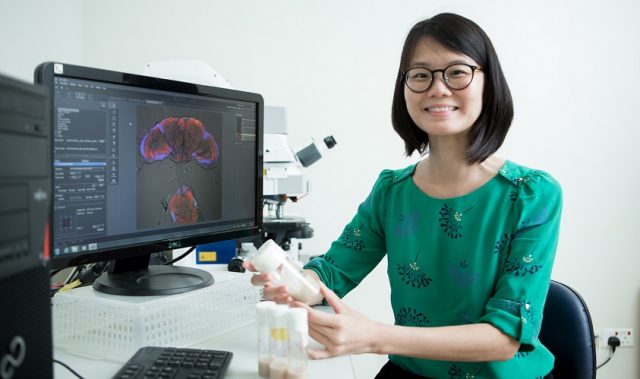
By designing real-time imaging techniques, the researchers were able to examine every cell within a pre-implantation mouse embryo without affecting its development. They observed differences in the way which certain proteins in each cell bind to their target genes, and how there were variances in cell behavior at every stage of the embryo’s development.
“Our findings as a result of this advanced technique have put forth a new paradigm of knowledge that would encourage more detailed microscopic analysis for future assisted reproduction procedures,” said Dr. Nicolas Plachta, a senior principal investigator at IMCB who led the study.
According to Plachta, further development of the real-time imaging technique may eventually enable fertility specialists to screen an embryo for genetic abnormalities without the need for physical manipulation.
The article can be found at: White et al. (2016) Long-Lived Binding of Sox2 to DNA Predicts Cell Fate in the Four-Cell Mouse Embryo.
———
Source: A*STAR; Photo: Nicolas Plachta.
Disclaimer: This article does not necessarily reflect the views of AsianScientist or its staff.