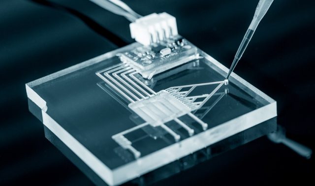
AsianScientist (Jan. 15, 2016) – Research teams from Seoul National University (SNU) and Chonnam National University (CNU) have developed a microchip that mimics bone tissues. The article was published in Lab on a Chip.
Their platform utilizes microfluidic technology that produces realistic cell culture microenvironments, similar in concept to an organ-on-a-chip. Organs-on-chips recreate the microarchitecture of a specific human organ and mimic their movements, and are generally developed as alternatives for animal testing in biomedical, pharmaceutical and toxicological areas.
One essential element for a successful outcome of tissue engineering—and specifically organs-on-chips—is to provide a vascular network that promotes the survival and proliferation of cells in any type of tissue or organ. In 2013, the SNU research group created a microfluidic device that could produce a perfusable and functional blood vessel. Their research suggested that this original vessel device could be a versatile platform for developing vascularized organs-on-chips.
For the recently developed microchip that mimics bone tissues, the SNU and CNU research teams, led by SNU Professor Jeon Noo Li from the Department of Mechanical Engineering, utilized the original vessel platform to develop new bone angiogenesis models.
Angiogenesis, the process of generating new blood vessels from pre-existing blood vessels, is involved in endochondral bone formation and bone fracture repair. In relation, the developed microchip predicts and reproduces physiological phenomena that occur in human and animal bone tissues. The result is a reproduction of highly porous and interconnected structures that mimic a real bone tissue matrix.
The SNU and CNU research teams explain in their paper of how the new bone angiogenesis model produces a more in vivo microenvironment for bone vessel sprouting. This development solves previous problems of the other bone tissue mimicking in vitro systems that have failed to reflect the complex chemical and mechanical microenvironment of the bone, and thus, were unable to provide similar in vivo conditions.
The microchip will offer a new approach for the investigation of complex biological phenomena, as well as for the analysis of drug responses and toxicities in bone tissues. Furthermore, the new model is expected to play a significant role in researching how to reproduce and regenerate new blood vessels.
The article can be found at: Jusoh et al. (2015) Microfluidic Vascularized Bone Tissue Model with Hydroxyapatite-incorporated Extracellular Matrix.
———
Source: Seoul National University; Photo: Yale Rosen/Flickr/CC.
Disclaimer: This article does not necessarily reflect the views of AsianScientist or its staff.