AsianScientist (Dec. 23, 2015) – Researchers have developed a genetic fluorescent probe that can label newly enlarged or generated synapses and then erase them when irradiated with blue light. The new genetic probe, described in Nature, could be a powerful tool for deepening understanding of brain function and learning in healthy or diseased individuals.
The development could also contribute significantly to understanding the mechanisms of dementia and post-traumatic stress disorder, according to the University of Tokyo researchers.
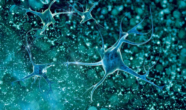
In the cerebral cortex several ten billions of neurons are connected by synapses forming elaborate neuronal networks. Notably, most excitatory glutamatergic synapses are formed on spiny protrusions that project out from neurons called dendritic spines, and the bigger dendritic spines there are the stronger synaptic connectivity.
Dendritic spines are dynamically modifiable through learning and memory resulting in changes in synaptic connectivity, and as a result are thought to be memory elements of the brain. However, this hypothesis has not been experimentally proven because there was no method to identify and alter large number of spines involved in memory formation.
Professor Haruo Kasai and his research group at the University of Tokyo Graduate School of Medicine’s Center for Disease Biology and Integrative Medicine have now developed optical probes which selectively label enlarged or newly generated spines, and, in addition, shrink and eliminate the labeled spines by blue laser irradiation to the living brain.
Since this probe is genetically encoded, it was incorporated into most neurons in the mouse motor cortex. In the experiment, mice were trained on a motor task to improve their performance, creating new dendritic spines and enlarging existing ones. Irradiating the motor cortex with blue laser light then eliminated learning-induced spine changes, resulting in reversal of the task performance and indicating that the spine changes are necessary for memory formation.
“This new technique allows us to visualize and control dendritic spines in action as the storage elements of memory for the first time,” said Kasai.
“We found that spines involved with memory formation were distributed densely in a relatively small fraction (10-20 percent) of all neurons in the cortex. This suggests that each memory is recorded in a specific strongly connected circuit,” Kasai added.
The ‘synaptic optogenetics’ probe developed by the group could be a powerful tool for further clarifying brain function in normal and disease states, according to the researchers.
The article can be found at: Hayashi-Takagi et al. (2015) Labelling and Optical Erasure of Synaptic Memory Traces in the Motor Cortex.
———
Source: The University of Tokyo.
Disclaimer: This article does not necessarily reflect the views of AsianScientist or its staff.