

AsianScientist (Jul. 09, 2026) – Osteoporosis is often called a “silent disease” because bones weaken gradually over many years without causing symptoms – until a fracture occurs. Detecting the condition early is crucial, but current clinical screening recommendations mainly focus on older women and other high-risk groups, which leaves some men, and younger adults with undiagnosed bone loss.
Osteoporosis affects over 200 million individuals globally and contributes to more than 8.9 million fractures annually. As populations age, especially in Asia where demographic transitions are accelerating, the incidence of osteoporosis-related fractures is projected to rise dramatically, with over 50 per cent of global hip fractures predicted to occur in Asia by 2050.
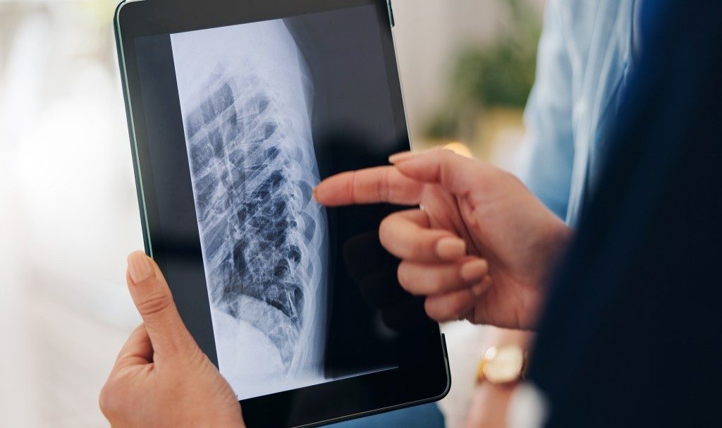
Now, a new study by researchers from National Taiwan University and St. Paul’s Hospital has shown that artificial intelligence (AI) can extract vital bone-health information from routine chest X-rays.
Published in npj Digital Medicine, the study demonstrates how AI can extract information about bone health from chest X rays that are already widely performed during routine health check-ups across Asia. The researchers state that this approach could help identify people who would not normally qualify for osteoporosis screening but who may nevertheless be at risk of fractures.
“This study demonstrates how artificial intelligence can transform existing healthcare workflows into scalable preventive-health strategies while supporting more equitable access to osteoporosis screening,” said Ray-E Chang, who is a co-corresponding author of the study, and a professor at the Institute of Health Policy and Management in National Taiwan University, Taipei.
Existing osteoporosis screening recommendations focus primarily on postmenopausal women, older adults and people with established clinical risk factors. As a result, younger adults, men and individuals with a healthy body weight often remain outside routine screening pathways. According to the study, this approach may overlook a substantial proportion of people with reduced bone density.
More than half of the participants who were confirmed to have abnormal bone density had a normal body mass index (BMI), highlighting a major blind spot in conventional risk-based screening.
Instead of replacing existing diagnostic methods, the AI system is designed to identify individuals who may benefit from further assessment using dual-energy X-ray absorptiometry (DXA), the current gold standard for measuring bone mineral density.
“Under Taiwan’s National Health Insurance system, we often rely on strict guideline-based criteria to decide who qualifies for DXA testing,” said Shu-Han Chen, MD, first author of the study, a family medicine physician, and leader of the Health Management Center at St. Paul’s Hospital in Taoyuan.
“Our findings suggest that AI-assisted chest X-ray analysis could help identify individuals who may otherwise be overlooked and who may benefit from confirmatory DXA testing,” Shu-Han added.
One of the key advantages of the approach is that it would repurpose imaging already being collected during routine medical care. Because no additional scans are required, opportunistic screening could be introduced with little extra cost or inconvenience for patients. By analysing existing images, healthcare providers may be able to identify early bone loss before fractures occur, enabling timely treatment and lifestyle interventions.
—
Source: t. Paul’s Hospital, Taiwan ; Image: The Yuri Arcurs Collection/magnific
You can find the study at: Advancing diagnostic equity through artificial intelligence chest radiograph screening for osteoporosis in Asian populations
Lead author: Shu-Han Chen, MD, family medicine physician, leader of the Health Management Center at St. Paul’s Hospital in Taoyuan, Taiwan.
Disclaimer: This article does not necessarily reflect the views of AsianScientist or its staff.