AsianScientist (Mar. 21, 2018) – A research group in Japan has discovered that immune cells are required to support brain development. They reported their findings in Nature Neuroscience.
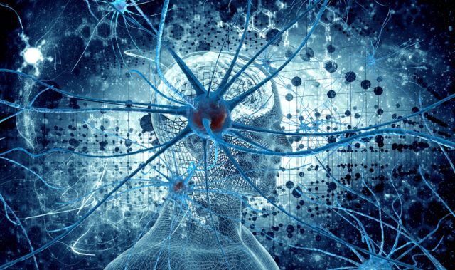
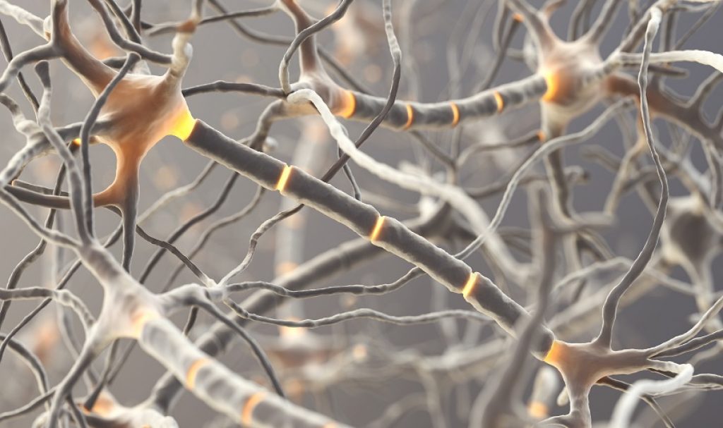
Neurons are specialized cells of the nervous system that communicate using electrical signals which propagate down long, wire-like projections called axons. The conduction of these signals requires myelin, a fatty substance that surrounds axons in much the same way that plastic coating surrounds an electrical wire.
Neurons are unable to produce their own myelin. Instead, supportive cells called oligodendrocytes are responsible for making the myelin sheath that wraps around axons. Several neurological disorders, including autism and schizophrenia, are thought to be driven in part by the failure of myelin to properly surround axons during development.
In the present study, researchers at Osaka University, Japan, have demonstrated that immune cells may play a key role in myelinating newly formed neurons. T- and B-cells are vital players in the immune system, circulating through the body, seeking infectious agents and mounting a protective response. These immune cells spend most of their time traversing the bloodstream and the lymph nodes but are thought to be blocked from accessing the brain.
“The central nervous system is protected from pathogens by a highly selective barrier that keeps the circulatory system physically separated from the brain,” said Assistant Professor Shogo Tanabe of Osaka University, who is the lead author of the study.
“It’s generally believed that this barrier also excludes immune cells from the brain. Our study suggests otherwise, though, as we found that a certain type of B-cell is quite abundant in the ventricles, meninges and choroid plexus in the brains of young mice. Even more surprising, these cells appear to stimulate axon myelination in the surrounding neurons,” he added.
The team discovered that a subset of B-cells, called B-1a cells, ensure that enough oligodendrocytes are available in the developing brain to support adequate myelination.
“We experimentally depleted B-cells from the brains of young mice and saw oligodendrocyte numbers drop significantly,” said Tanabe. “It turns out that so-called natural antibodies secreted by B-1a cells induce oligodendrocyte precursors to proliferate. These antibodies are typically involved in immune surveillance, but in this case they promote the myelination of axons.”
Recent studies have shown that T-cells can occupy the meninges and play a role in learning and memory. However, according to the researchers, this is the first study implicating B-cells in myelin production during early development. The study may have broad implications for diseases driven by early defects in neuronal growth.
“Prior work has indirectly hinted at a role for B-cells in neurodevelopmental disorders,” said Professor Toshihide Yamashita of Osaka University who is the senior lead author of the paper.
“Our findings provide direct evidence that B-cells reside in the mouse neonatal brain and promote both oligodendrocyte proliferation and neuron myelination. This suggests to us that B-cell dysfunction in early development may contribute to later mental disorders, a possibility that we believe deserves further exploration in future studies.”
The article can be found at: Tanabe et al. (2018) B-1a Lymphocytes Promote Oligodendrogenesis During Brain Development.
———
Source: Osaka University; Photo: Shutterstock.
Disclaimer: This article does not necessarily reflect the views of AsianScientist or its staff.