AsianScientist (Feb. 19, 2018) – Scientists in Singapore have developed a method to grow patient-derived liver tumor organoids for drug testing. Their findings are published in Biomaterials.
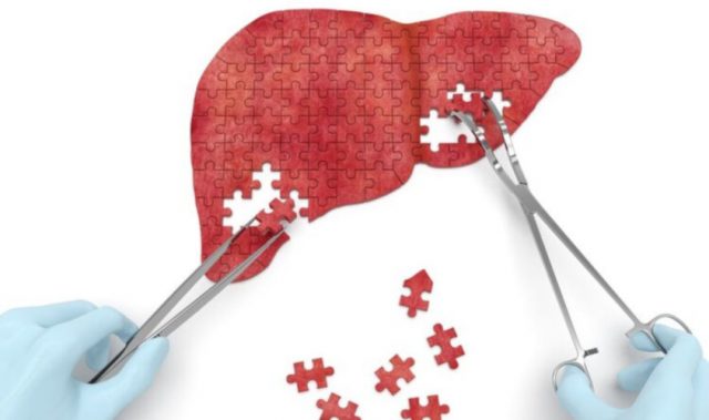
Liver cancer is one of the top causes of cancer deaths in the world, with a dearth of approved treatments. A major challenge in developing effective drugs for liver cancer is that current preclinical tumor models do not accurately replicate features of the tumor and the tumor environment in humans, causing many potential drugs to fail in clinical testing.
To more accurately mimic these features, researchers have developed models of liver tumors called patient-derived xenografts (PDX). Although these models provide a truer picture of how effective potential cancer drugs would be in humans, they are also expensive and time-consuming to create. Growing these PDX cancer cells in culture would be more cost effective for drug screening. However, so far, attempts to culture these cells fail to reflect the three-dimensional (3D) tumor structure and the tumor environment.
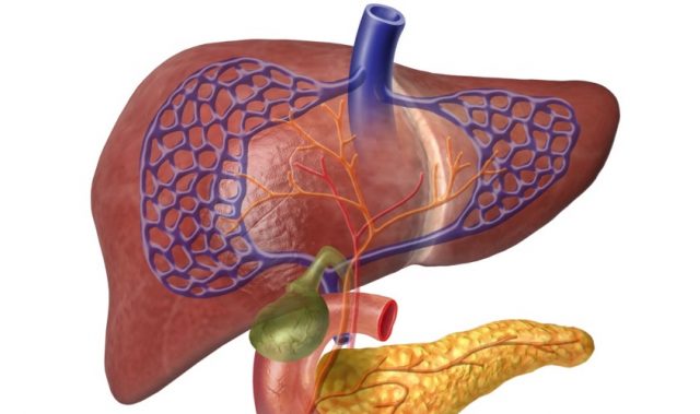
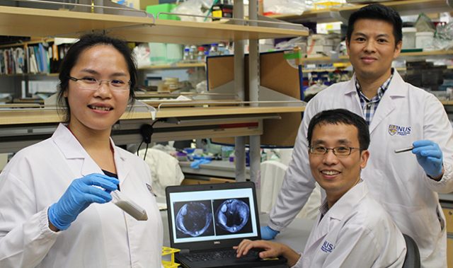
In this study led by Dr. Eliza Fong and Dr. Toh Tan Boon of the National University of Singapore (NUS), the researchers grew patient-derived liver cancer cells from 14 patients on synthetic 3D scaffolds made of a plant-based porous hydrogel. The researchers engineered the spongy scaffolds with optimized biochemical and mechanical properties to help the liver cancer cells maintain their proper shape and function and grow as organoids.
After growing the organoids for one to three weeks, the team verified that the liver cancer cells in the organoids were alive and growing. Liver cancer cells usually contain specific genetic changes that are absent in normal liver cells. Most of the organoids retained the same key genetic changes that were in the source PDX cells, suggesting that they accurately mirror the disease condition in their respective patients.
The organoids also maintained an important feature of liver tumors called intra-tumor heterogeneity, in which distinct populations of liver cancer cells were present within the same tumor and affected the tumors’ responses to treatment. This feature is one advantage of using organoids over traditional cell culture methods in which all cells are identical.
Another attractive feature is the small size of the 3D scaffolds containing the organoids. They can easily fit inside the well of a 96-well microtiter plate, a standard platform for high-throughput drug screening, which enables many drugs to be tested at the same time.
With this technology, one PDX can be used to produce tens to hundreds of such organoid-containing scaffolds. Combined with their ability to recapitulate the genetic features and heterogeneity of the original liver tumors, these tumor avatars have the potential to revolutionize the screening and development of liver cancer drugs.
“This study truly epitomizes the positive synergy we can achieve in growing patient tumors outside the body by marrying advances in tissue engineering with cancer biology,” said Fong.
“Having a reliable platform to grow liver cancer patient-derived cells is a major step in personalized medicine as we can now use them for increased throughput drug sensitivity testing,” Toh added.
The article can be found at: Fong et al. (2018) Generation of Matched Patient-derived Xenograft in vitro-in vivo Models using 3D Macroporous Hydrogels for the Study of Liver Cancer.
———
Source: National University of Singapore.
Disclaimer: This article does not necessarily reflect the views of AsianScientist or its staff.