AsianScientist (Sep. 29, 2017) – Scientists in Korea have devised a method to directly observe the movement of molecules without the need for dyes. They report their findings in Advanced Materials.
Moving, vibrating and leaping molecules make up our world. Capturing their movement is not an easy task; to observe molecular motion, scientists typically need to stain samples with metal or dye molecules to make them visible inside a graphene pocket during electron microscopy.
Metals have high reflexibility, making them useful for getting good images. However, the chemical bonds between the sample and the metal or dye changes the characteristics of the sample molecule.
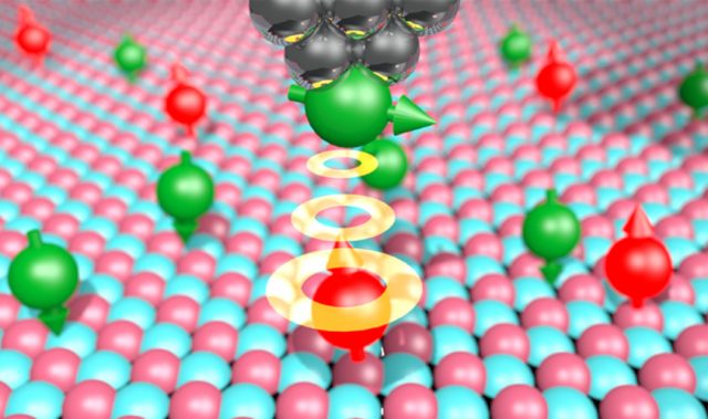
In this study, scientists at the Center for Soft and Living Matter, within the Institute for Basic Science (IBS), were able to see the movement of molecules stored inside a graphene pocket without the need to stain them. They visualized chains of atoms, called polymers, swimming in a liquid inside graphene pockets. These pockets consist of three to five graphene layers on the bottom and two on top. Because of how thin the layers are, the contents within the graphene pockets can be observed in real time without any staining.
More importantly, the sheets are impermeable to small molecules and prevent the electron beam from instantly harming the sample—the scientists had an average of 100 seconds to watch the dynamic movement of individual polymer molecules before the molecules were destroyed by the electron beam. During these valuable seconds, molecules changed position, rearranged or ‘jumped around.’
“It was amazing to watch these flexible organic macromolecules dancing around,” said Dr. Hima Nagamanasa of IBS who was the first author of the paper. “Molecules move much more quickly in bulk. We were surprised to see that they are moving more slowly here. We believe that attachment to the surface of the pocket worked in our favor to slow them down. Otherwise, we would probably have just seen a blurred image.”
The scientists worked with two polymers: one containing sulfur (polystyrene sulfonate) and the other without (polyethylene oxide). This allowed them to show that the contrast under the microscope was derived from the polymer structure—made of carbon and hydrogen atoms—rather than from the sulfur.
“Most molecules produced by living organisms have a backbone made of carbon and hydrogen, and that’s why we hope to extend this research to the study of interactions between DNA and proteins,” explained Dr. Wang Huan of IBS who was a co-author of the study.
The article can be found at: Nagamanasa et al. (2017) Liquid-Cell Electron Microscopy of Adsorbed Polymers.
———
Source: Institute for Basic Science; Photo: Shutterstock.
Disclaimer: This article does not necessarily reflect the views of AsianScientist or its staff.