AsianScientist (May 17, 2016) – Scientists in Japan have shown that the algae Chlamydomonas reinhardtii relies on organs known as eyespots to move towards light sources and can use its cell body as a convex lens to focus light. Their findings have been published in Proceedings of the National Academy of Sciences.
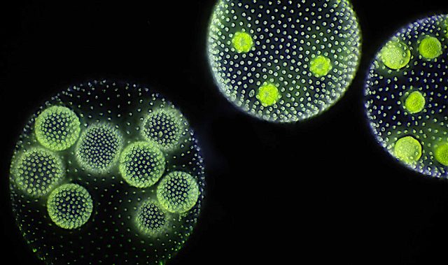
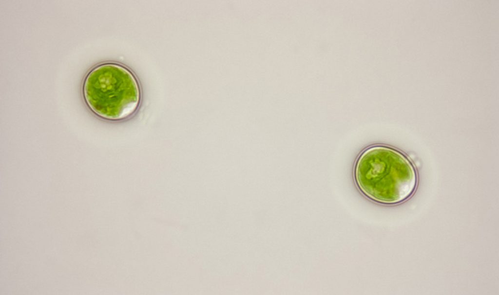
C. reinhardtii is a unicellular green algae that lives in fresh water throughout the world. It is well known for moving aqueous environments by beating flagella, a hair-like organelle that generate force for swimming.
Notably, the cells can change their swimming directions upon sensing light; a response referred to as phototactic behavior. The algae swim and find locations in their environments for optimizing photosynthesis by either moving towards light (positive phototaxis) or away from it (negative phototaxis).
However, in spite of their widespread use as a model organism for phototactic behavior, the role of pigments in the subcellular organelle called eyespots in determining the direction of cell movement relative to light sources is still unclear.
Now, the research team, led by Professor Ken-ichi Wakabayashi from Tokyo Institute of Technology, has identified a mutant which has no eyespots and a reversed responsiveness to light, moving in the opposite direction from wild type C. reinhardtii strains.
To explain these findings, the researchers designed experiments to determine whether the cells behaved as lenses to focus and condense light onto photoreceptors that are located on the equatorial region of the surface of one side of the algae.
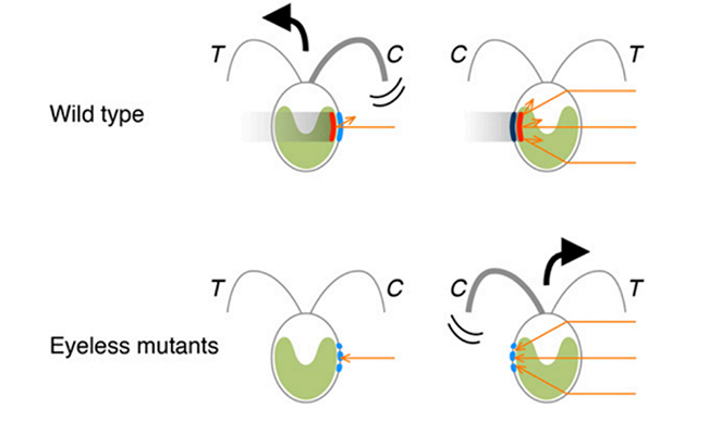
In normal wild type cells, the eyespot pigment is located below the photoreceptors. When the eyespot is facing a light source, the light signal is reflected and amplified by the pigment layers constituting the eyespot onto the photoreceptors. On the other hand, when the eyespot and photoreceptors are pointing in the opposite direction to the light source, the light does not reach the photoreceptors because it is shielded by the eyespot pigments, and the cell does not change its swimming direction.
Furthermore, in the case of mutant cells without eyespot pigments, when the photoreceptors are pointing directly towards a light source, the light signal falls directly onto the photoreceptors but it is too weak to trigger turning of the cells. In the opposite orientation, with the photoreceptors are pointing away from the light source, the light signal is focused and condensed by the lens-action of the cell onto the photoreceptors, leading to the turning of Chlamy.
The eyespot pigments therefore play a crucial role in determining the sign of phototaxis, by shielding the photoreceptors from light condensed by the cellular lens onto the back of the eyespot, according to the researchers.
The article can be found at: Ueki et al. (2016) Eyespot-dependent Determination of the Phototactic Sign in Chlamydomonas reinhardtii.
———
Source: Tokyo Institute of Technology; Photo: Marc Perkins/Flickr/CC.
Disclaimer: This article does not necessarily reflect the views of AsianScientist or its staff.