AsianScientist (Nov. 12, 2015) – In a study published in the journal Angewandte Chemie International Edition, a team of chemists and biologists at the Institute of Transformative Bio-Molecules (ITbM), Nagoya University has developed a fluorescent dye with enhanced photostability to enable continuous live cell imaging by STED microscopy. Called C-Naphox, it opens doors to observe real-time biological events for extended time periods with high resolution.
Bioimaging by fluorescence microscopy is a useful technique to study the localization and movement of molecules in living cells by fluorescence. Yet, the gradual degradation of fluorescent dyes when exposed to the high intensity light necessary for super resolution microscopy has been a major obstacle for long-term observations.
A photostable fluorescent dye for super resolution microscopy could serve as a powerful tool to visualize biological events and structural details in living cells at real-time for prolonged recording periods.
Advances in super-resolution techniques such as stimulated emission depletion (STED) microscopy (2014 Nobel Prize in Chemistry) have enabled nanoscale visualization of biological systems. STED microscopy allows up to ten times higher resolution in living cells with respect to conventional microscopes by employing a fluorescence excitation beam in combination with a surrounding doughnut-shaped STED beam. While the molecules at the center are excited to a higher energy state and fluoresce, the molecules exposed to the STED beam are confined to the ground energy state.
However, the light intensity required for super-resolution fluorescence microscopy is much higher than conventional microscopy, which leads to photodegradation of fluorescent dye molecules. As the spatial resolution of STED imaging correlates with an increase in STED light intensity, photodegradation of fluorescent dyes becomes a serious issue.
Representative photoresistant fluorescent dyes such as Alexa Fluor® 488 and ATTO 488 are also known to encounter photodegradation in STED microscopy, making it difficult to conduct continuous live-imaging of biological systems while retaining high resolution.
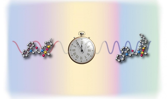
To overcome this issue of reduced resolution in STED imaging due to photodegradation, ITbM’s team led by Principal Investigators Shigehiro Yamaguchi, a synthetic chemist and Tetsuya Higashiyama, a plant biologist have developed a fluorescent dye, C-Naphox that has enhanced photostability relative to conventional dyes. C-Naphox has demonstrated to be extremely photoresistant with almost no degradation of fluorescence even after prolonged STED imaging in live cells.
C-Naphox (diarylmethylene-bridged naphthophosphole P-oxide) consists of an aromatic framework with an amino moiety incorporated for its electron-donating properties and phosphorus oxide for its electron-accepting properties, leading to intense fluorescence emissions.
“Although the previously synthesized molecules in the Yamaguchi group have also led to high fluorescence intensity, it is the carbon-bridged structure in C-Naphox that is the key to its extremely high resistance to high intensity light,” says Aiko Fukazawa, an associate professor in Yamaguchi’s group.
Together with Masayasu Taki, an associate professor and Wang Chenguang, a postdoctoral research fellow in Yamaguchi’s group, they optimized the structure of the fluorescent dye for live cell imaging.
“C-Naphox and other derivatives were rapidly synthesized by Dr. Wang in less than ten steps and were found to be air-stable for months at room temperature,” says Fukazawa. “Furthermore, C-Naphox exhibited no significant toxicity towards cells at micromolar concentrations that are usually required for cell staining.”
The photostability of the newly synthesized dye was proven by irradiation experiments with a xenon lamp. The relative absorbance of C-Naphox was compared to that of representative commercially available STED imaging fluorescent probes, Alexa Fluor® 488 and ATTO 488.
The results of the experiments showed that over 99 percent of C-Naphox remained intact even after 12 hours of irradiation, whereas only 26 percent of Alexa Fluor® 488 remained present after two hours. Although 97 percent of ATTO 488 persisted after two hours of irradiation, the amount present was reduced to 59 percent after 12 hours.
HeLa cells were then stained by either C-Naphox or Alexa Fluor® 488 and exposed to repeated STED imaging conditions. While Alexa Fluor® 488-stained cells showed a decrease in fluorescence signal intensity after five recordings, the intensity of C-Naphox remained virtually unchanged under the same conditions.
“As STED microscopy is an extremely powerful tool to visualize extremely small structures, we hope to apply these types of photoresistant molecules to study biological events such as the formation of actin filaments in cells,” said Taki.
“C-Naphox is an extremely photoresistant fluorescent dye that allows repeated STED imaging that has been difficult with conventional dyes. We are currently conducting further optimization studies in order to improve the practicality of C-Naphox as a useful fluorescent probe in continuous time lapse live cell STED imaging and multi-color imaging,” said Fukazawa.
The article can be found at: Wang et al. (2015) A Phosphole Oxide Based Fluorescent Dye with Exceptional Resistance to Photobleaching: A Practical Tool for Continuous Imaging in STED Microscopy.
———
Source: Institute of Transformative Bio-Molecules.
Disclaimer: This article does not necessarily reflect the views of AsianScientist or its staff.