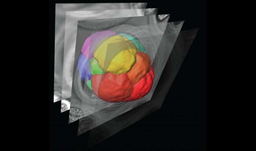
AsianScientist (Sep. 3, 2015) – A collaboration between biologists and engineers at Monash University has led to the development of a new non-invasive image processing technique to visualize embryo formation. Researchers were able to see, for the first time, the movement of all of the cells in living mammalian embryos as they develop under the microscope.
This advancement has important implications for in vitro fertilization (IVF) treatments and pre-implantation genetic diagnosis (PGD). In the future, this approach could help with embryo selection before the embryo is implanted back into the uterus to improve IVF success rates.
The research, published in Developmental Cell, provides new insights into embryo formation and challenges the prevailing model of cell placement through division.
Mammalian embryos start out as a small group of identical cells. Then at an early stage, some of these cells take up an internal position within the embryo. These internal cells are the ones that will go on to form all of the cells of the body while the remaining outer cells go on to form other tissues such as the placenta.
For many years, researchers hypothesized that the internal cells adopt their position through a special process of cell division, but due to technological limitations, this had never actually been shown. Using the newly developed imaging methods, the researchers were able to demonstrate that this model of embryo formation was incorrect.
The researchers then applied cutting-edge laser techniques previously used in fly and plant embryos or cultured cells only to the mammalian embryo to determine what forces were acting on the cells to make them move inside the embryo. They were able to see how the cells moved and changed shape over time as they were ‘pushed’ inside to form the internal mass.
They showed that there are differences in the tension of the membranes of the cells and these differences are what determine which cells will move inside to form the body. By altering the tension of the cells using lasers or genetic manipulations, researchers could change which cells move inside the embryo.
These findings also offer future potential to make alterations to improve inter-cellular forces and cell formation.
The article can be found at: Samarage et al. (2015) Cortical Tension Allocates the First Inner Cells of the Mammalian Embryo.
———
Source: Monash University.
Disclaimer: This article does not necessarily reflect the views of AsianScientist or its staff.