AsianScientist (Sep. 9, 2014) – Scientists now understand how thalamocortical (TC) neurons shift from burst to tonic mode, thanks to research that has been published as the cover article in The Journal of Neuroscience.
Neurons sense a signal through branching dendrites, carry this signal to the cell body, and send it onwards through a long axon to signal the next neuron. However, neurons can function in many different ways; some of which researchers are still exploring. Some signals that the dendrites receive do not continue to the next neuron; instead they seem to change the way that the neuron handles the subsequent signals. These non-transmitted signals are thought to help neurons function as part of a large network, although the precise mechanism is not known.
Dr. Sigita Augustinaite, a researcher in the Optical Neuroimaging Unit at the Okinawa Institute of Science and Technology Graduate University (OIST), has now proposed a mechanism explaining how neurons help the network function.
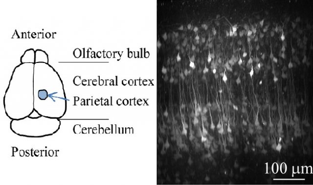
Dr. Augustinaite studies the visual pathway, where signals from the retina are sent to the visual cortex, where the brain interprets signals from the eye. Between the eye and the visual cortex, the signals must pass through the visual thalamus, that is, through the TC neurons. These neurons can switch between a “sleeping” state and a “waking” state depending on input they receive from neurons and other brain areas. When an animal is awake, TC neurons transmit the incoming retinal signals on to the cortex, but when the animal is asleep, the neurons block retinal signals.
To understand what regulated the switch between sleeping and waking states, she conducted experiments in acute brain slices, small pieces of brain tissue where neurons stay alive and maintain their physiological properties. Firstly, she added glutamate to dendrites far from the cell body to emulate a feedback signal from the visual cortex. Then she measured the neuron’s response, shown as a voltage difference between inside and outside of the membrane.
Dr. Augustinaite found that stimulating the neurons in this way depolarizes their membranes, creating something called NMDA spike/plateau potentials. If strong enough, depolarization can cause a neuron to fire an action potential, which travels through the axon to activate other neurons. But if NMDA spike/plateaus induces action potentials, signals from the cortex and signals from the retina would be indistinguishable.
With her experiments, Dr. Augustinaite showed that the NMDA spike/plateau potentials in TC neurons do not trigger action potentials. Instead, they lift the voltage of the membrane, changing the neuron’s properties for few hundred milliseconds, creating conditions for reliable signal transmission from retina to cortex.
“The research gives, for the first time, a clear view on what dendritic potentials are good for,” explained Professor Bernd Kuhn, who leads the lab where Dr. Augustinaite works. “It points directly to the mechanism,” he concluded.
“This mechanism could also be used in many other neuronal circuits, where one input regulates how another input moves through the network,” Dr. Augustinaite said. “This mechanism is an exciting logical element in the neuronal network, but just the start of putting the puzzle together.”
The article can be found at: Augustinaite et al. (2014) NMDA Spike/Plateau Potentials in Dendrites of Thalamocortical Neurons.
——-
Source: Okinawa Institute of Science and Technology Graduate University.
Disclaimer: This article does not necessarily reflect the views of AsianScientist or its staff.