AsianScientist (Jul 15, 2014) – Scientists have developed a non-invasive method of detecting circulating tumor cells that can be combined with genetic screening. This research has been published in the journal Gut.
Taking a biopsy of a tumor from a cancer patient requires surgery that may not be well tolerated in the elderly or weak. Instead of biopsies, it is possible to study a patient’s cancer by simply collecting a blood sample and looking for circulating tumor cells (CTCs), cells from the tumor that have been shed into the bloodstream.
It is challenging to detect CTCs because there are only very small quantities of CTCs in the bloodstream. Currently, the CellSearch system is widely used to detect CTCs, where antibodies are used to target the major epithelial cell surface marker known as the epithelial cell adhesion molecule. However, recent research has shown that heterogeneous CTCs, which have both epithelial and mesenchymal characteristics, exist, highlighting the need for CTC capture systems that are able to detect CTCs irrespective of the epithelial cell marker.
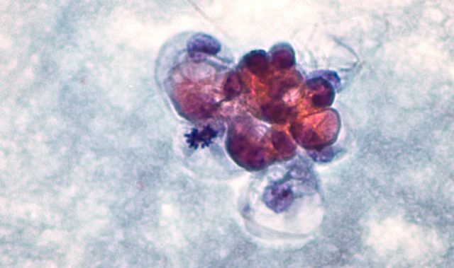
To overcome these problems, the research team led by Professor Toshiyoshi Fujiwara from Okayama University Graduate School of Medicine, Dentistry, and Pharmaceutical Sciences exploited the high telomerase activity of malignant tumor cells. Using a modified adenovirus (OBP-401) which only replicates in cancer cells, they were able to label the CTCs with green fluorescent protein (GFP). This allowed them to capture the extremely low quantities of live CTCs from the millions of background blood leukocytes.
By combining OBP-401-based CTC capture system and genetic testing, the research team was able to detection of KRAS and BRAF mutations in blood samples taken from patients with colorectal cancer. Notably, these mutations were determined to be identical to the ones seen in the primary tumours of the patients.
Targeted cancer treatment for individual patients is currently carried out by analysis of primary tumors. The difficulties with this approach are that the primary tumors contain very few cells that cause metastasis, and samples for analysis are obtained by needle core biopsies or surgical removal of tumor tissue, invasive procedures that may not be possible in certain regions such as the brain.
“This ‘liquid biopsy’ via a simple blood test could be carried out in real time and enables optimized and timely decisions for therapeutic intervention,” stated the researchers.
The article can be found at: Shigeyasu et al. (2014) Fluorescence virus-guided capturing system of human colorectal circulating tumour cells for non-invasive companion diagnostics.
——
Source: Okayama University.
Disclaimer: This article does not necessarily reflect the views of AsianScientist or its staff.