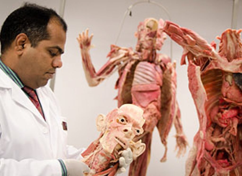
AsianScientist (May 27, 2013) – Nanyang Technological University’s (NTU) new medical school will be pioneering the use of plastinated bodies for its anatomy classes.
Plastinated bodies are human bodies which people have pledged towards the learning and advancement of science upon their death and are preserved through plastination. These human bodies are preserved by replacing the fat and water in body tissues with plastic.
The human bodies and body parts, known as specimens, will be used in anatomy classes taught at the Lee Kong Chian School of Medicine (LKCMedicine), and will be ready for the first batch of 54 medical students this August.
The new medical school, jointly set up by NTU and Imperial College London, had ordered the human bodies and specimens from Germany, which were prepared to the school’s specifications. There are two whole bodies and various body parts such as hearts, lungs, brains, limbs, and torsos.
With the use of plastinated bodies and specimens, traditionally preserved cadavers, which are in short supply in Singapore, will no longer be needed by LKCMedicine. Aside from that, there are many distinct advantages of using plastinated specimens over traditional cadavers, says Assistant Professor Dinesh Kumar Srinivasan, the Lead for Anatomy Teaching at LKCMedicine.
“These highly detailed, plastinated human specimens are very durable and can be repeatedly handled by students without deterioration and it can be stored just like any inert object,” Prof Srinivasan said. “They may be used in a much broader range of educational settings, since we no longer have to take extra steps to re-preserve the body in embalming chemicals, as we would need to for traditional cadavers each time they are used in class.
“In addition, plastinated human specimens can be used to demonstrate difficult structures and dissection areas in high-definition, such as the blood vessels in the brain or the nerves in the spine. Such intricate details, essential in learning anatomy, will start degrading in traditionally preserved cadavers after repeated use.”
Students at LKCMedicine will be using the plastinated human bodies for anatomy classes in their first and second year at the school.
“The anatomy lab experience will be very different for a third-year student as compared to a first-year medical student. First-year students will be mostly focused on identifying structures from their textbook, whereas third-year student will be interested in how the anatomy has clinical value and how it relates to real life patients,” he added.
In addition to plastinated human bodies, students will also learn from microscopic slides to understand the microscopic anatomy of cells and tissues of humans. Anatomy classes will be also supplemented by the use of the Anatomage Table, which displays life-sized 3D images of full body anatomy using computed tomography (CT) and magnetic resonance imaging (MRI) scans.
——
Source: NTU.
Disclaimer: This article does not necessarily reflect the views of AsianScientist or its staff.