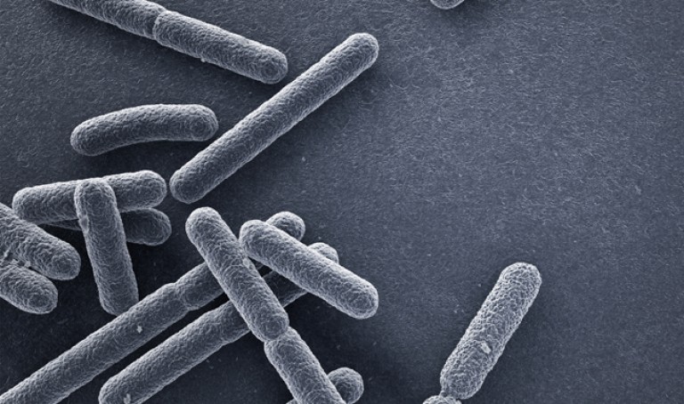
AsianScientist (Apr. 4, 2016) – Professor Park YongKeun of the Physics Department at the Korea Advanced Institute of Science and Technology and his research team have developed a powerful method for 3D imaging of live cells without staining. Their 2D/3D/4D holographic microscope, named holotomography (HT)-1, allows for real-time label-free visualization of biological cells and tissues.
The researchers have announced the launch of their new microscopic tool to the global marketplace through a start-up that Park co-founded, TomoCube.
Current fluorescence microscopy techniques require the use of externally-derived labeling agents to render high-contrast molecular information. Therefore, possible drawbacks include photo-bleaching, photo-toxicity and interference with normal molecular activities. In particular, it is particularly difficult to use fluorescent microscopy with immmune or stem cells that need to be reinjected into the body.
The HT-1 is an optical analogy of X-ray computed tomography (CT). Both HT and X-ray CT share the same physical principle—the inverse of wave scattering. The difference is that HT uses laser illumination, whereas X-ray CT uses X-ray beams.
From the measurement of multiple 2D holograms of a cell, coupled with various angles of laser illuminations, the microscope can reconstruct the 3D refractive index (RI) distribution of the cell. The reconstructed 3D RI map provides structural and chemical information of the cell including mass, morphology, protein concentration and dynamics of the cellular membrane.
The HT-1 also enables users to quantitatively and non-invasively investigate the intrinsic properties of biological cells; for example, dry mass and protein concentration.
“As one of the two currently available, high-resolution tomographic microscopes in the world, I believe that the HT-1 is the best in class regarding specifications and functionality. Users can see 3D/4D live images of cells without fixing, coating or staining cells. Sample preparation times are reduced from a few days or hours to just a few minutes,” said Park.
“Our technology has set a new paradigm for cell observation under a microscope. I expect that this tomographic microscopy will be more widely used in future in various areas of pharmaceuticals, neuroscience, immunology, hematology, and cell biology.”
———
Source: KAIST; Photo: Tomocube.
Disclaimer: This article does not necessarily reflect the views of AsianScientist or its staff.