AsianScientist (Mar. 21, 2018) – A team of scientists from the Li Ka Shing Faculty of Medicine of The University of Hong Kong (HKU) and Imperial College London has developed a way to see through human brain tissue at high resolution. They published their findings in Nature Communications.
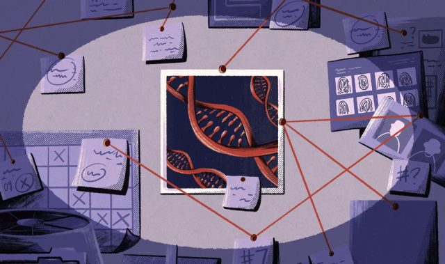
To understand how the brain works, scientists need to map how nerve cells (neurons) are wired to form circuits in both healthy and disease states. Traditionally, this was accomplished by cutting brain tissue into thin slices and tracing the nerve fibres over many sections.
However, this approach is difficult and labor-intensive as the neuronal circuitries span across great distances in three dimensions (3D) and are tightly entangled microscopically. To avoid the sectioning of tissues, tissue clearing techniques—methods that turn opaque tissue transparent—have been developed, enabling high resolution and deep imaging of neuronal circuitries.
Although such techniques have been very effective on rodent brain tissue, only limited studies have found success with human brain tissue. The difficulties and challenges may be attributed to fundamental differences between the human and the mice brain.
To overcome these barriers, the authors of this study developed a tissue clearing solution, which they called OPTIClear. The solution selectively adjusts the optical properties of tissue without damaging or changing their structural components.
Combined with fluorescent staining and other tissue processing methods, OPTIClear is a simple yet versatile tool for the study of microscopic structures in the human brain. Nerve cells, glial cells, and blood vessels were visualized in exquisite detail, with their 3D relationship determined.
For example, the team performed 3D morphological analysis on human brainstem dopaminergic neurons in the millimetre scale and imaged more than 3,000 large neurons in the human basal forebrain in merely five days, a normally extremely laborious task that takes at least three weeks. These neurons have been implicated in neurological and psychiatric diseases such as dementia and depression. The researchers noted that OPTIClear can also be applied in both archived (>30 years) and clinical specimens.
“We hope that a better understanding of the connections and circuitries of the brain will help uncover the pathologies that underlie the common degenerative diseases of the brain, such as Alzheimer’s and Parkinson’s disease,” said Professor Wu Wutian, Honorary Professor at the School of Biomedical Sciences in HKU, who supervised the study.
“In principle, this method is also applicable to other human organs and clinical specimens. We hope that this technique can be used in the study of other diseases, as well as help us to unravel the mysteries of the human body,” said Mr. Lai Hei-ming of HKU, who is the first author of the study.
The article can be found at: Lai et al. (2018) Next Generation Histology Methods for Three-dimensional Imaging of Fresh and Archival Human Brain Tissues.
———
Source: The University of Hong Kong; Photo: The University of Hong Kong.
Disclaimer: This article does not necessarily reflect the views of AsianScientist or its staff.