AsianScientist (Jun. 5, 2017) – Researchers have invented a medical gamma-ray detector that can take high-resolution, multicolor 3D images in live mice. Furthermore, their device described in a study published in Scientific Reports weights only 580 g and fits in the palm of a hand, making it the world’s most compact Compton camera.
Conventional radiological images provide only black and white figures in 2D space. The situation is basically the same for single photon emission tomography (SPECT) and positron emission tomography (PET), which are the two most common molecular imaging techniques used in nuclear medicine.
PET is used especially for early cancer and Alzheimer’s disease detection, but radioactive tracers suitable for each detector are limited in terms of energy. For example, PET can only image monochromatic gamma rays thus provide black and white 2D images. Moreover, production of PET tracers, usually made by a cyclotron facility in medical centers, is inevitably costly.
“All of these problems could be addressed if gamma rays of arbitrary energy could be easily visualized in 3D space,” said Jun Kataoka, professor of applied physics at Waseda University. “This would be as revolutionary as black and white television turning into color, dramatically increasing the amount of information we could obtain from an image.”
Although SPECT and PET are clinically used, the radioactive tracers suitable for each detector have been limited. SPECT only images low-energy gamma rays less than 400 kilo-electron volts (keV), and PET can image only positron emitting sources of 511 keV. A Compton camera, which can image energy from a few hundred keV to more than mega-electron volts (MeV), would give clinicians more imaging options.
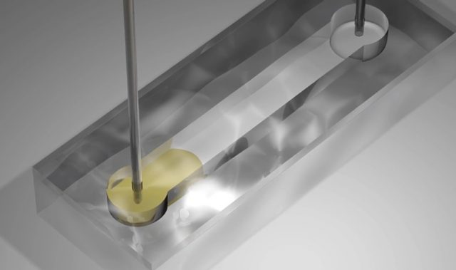
The Compton camera developed by Kataoka’s group has a high detection efficiency and practical spatial resolution, enabling flexible measurements. By rotating the camera around a mouse that had been given three different radioactive tracers—iodine (131I), strontium (85Sr), and zinc (65Zn)—the researchers were able to simultaneously image all three traces almost in real-time with a resolution of 3mm, comparable to PET.
They discovered that the tracers iodine, strontium and zinc accumulated in the thyroid, bones and liver respectively, confirming that these new tracers concentrated in each target organ.
“The measurement time took 10 minutes per angle, so we were able to obtain an image taken from 12 angles in just two hours. The time could be reduced even more by using multiple Compton cameras. For example, if there are 12 Compton cameras surrounding an object, the same image as this study could be obtained in just 10 minutes, suggesting a new way to understand biodynamics by looking at how a drug is taken into the body in 10-minute increments.”
Based on this study, Kataoka is now working towards developing a gamma-ray camera which works like the human eye.
“The human eye can instantly distinguish the colors and brightness of light from all directions, as well as determine the object’s shape in 3D from the displacement between the left and right eye. Therefore, stereoscopic imaging becomes theoretically feasible by using multiple ultra-compact Compton cameras.”
Though not limited to the medical field, this technology could help track behaviors of cancer cells and minerals in the body by combining the conventional PET drugs with newly found tracers, calculate the survival rate of a transplanted organ, develop cheaper and more convenient drugs for medical imaging, and monitor online the effectiveness of particle therapy by measuring various prompt gamma rays emitted during treatment.
“As radiation technology is still emerging, we look forward to expanding the possibilities of next-generation radiation imaging with this ‘on demand’ Compton camera,” Kataoka said.
The article can be found at: Kishimoto et al. (2017) First Demonstration of Multi-color 3-D in vivo Imaging Using Ultra-compact Compton Camera.
———
Source: Waseda University.
Disclaimer: This article does not necessarily reflect the views of AsianScientist or its staff.