AsianScientist (May 31, 2017) – In a study published in Nature Communications, researchers at the Korea Advanced Institute of Science and Technology (KAIST) describe how they have used a holographic technique to control the position, orientation and shape of oddly shaped microscopic samples.
Conventional optical manipulation techniques called ‘optical tweezers’ are an invaluable tool for manipulating microscopic particles in 3D. Using a tightly-focused laser whose beam diameter is smaller than one micrometer (1/100 of hair thickness), optical tweezers can generate an attractive force on microscopic particles moving toward the beam focus.
However, most experiments using optical tweezers have only trapped spherical particles for which the responding motion can be easily predicted. Conventional optical tweezers induce unstable motions of particles with more complex shapes, hindering their use on microscopic objects with complex shapes such as living cells.
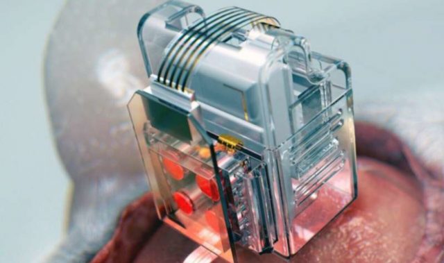
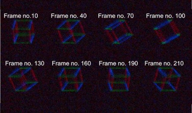
In the present study, researchers have developed a new optical manipulation technique that can trap complex objects of arbitrary shapes. This technique first measures 3D structures of an object in real time using a 3D holographic microscope, which shares the same physical principle of X-ray computed tomography (CT) imaging.
Based on the measured 3D shape of the object, the researchers precisely calculated the shape of light that can stably control the object. When the shape of light is the same as the shape of the object, the energy of the object is minimized, making the trapping stable.
Moreover, by controlling the positions, directions and shape of the light, it is possible to control the motion of a microscopic object in 3D space and change its shape. Since this process resembles the generation of a mold for casting a statue having desired shape, the researchers named their technique tomographic mold for optical trapping or TOMOTRAP.
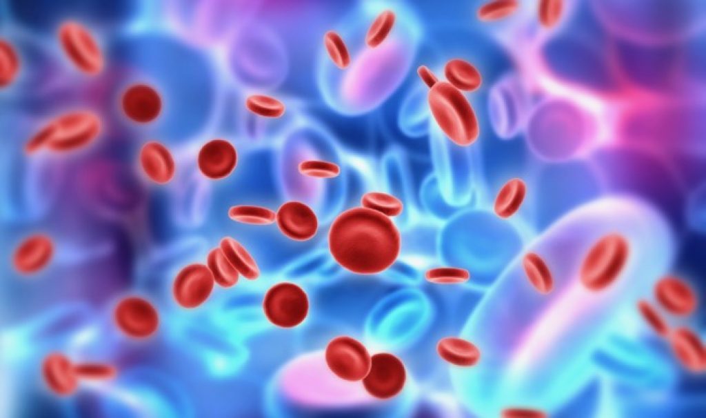
Using TOMOTRAP, the team succeeded in trapping individual human red blood cells stably, rotating them with desired orientations, folding them in an L-shape, and assembling two red blood cells together to form a new structure. In addition, colon cancer cells having a complex structure could be stably trapped and rotated at desired orientations.
“Our technique has the advantage of controlling the 3D motion of complex shaped objects without knowing prior information about their shape and optical characteristics, and can be applied in various fields including physics, optics, nanotechnology, and medical science,” said study co-author Professor Park YongKeun.
Dr. Kim Kyoohyun, the lead author of the study, noted that this technique can induce controlled deformation of biological cells with desired shapes.
“This approach can be also applied to real-time monitoring of surgical prognosis of cellular-level surgeries for capturing and deforming cells as well as subcellular organelles,” he added.
The article can be found at: Kim & Park (2017) Tomographic Active Optical Trapping of Arbitrarily Shaped Objects by Exploiting 3D Refractive Index Maps.
———
Source: Korea Advanced Institute of Science and Technology.
Disclaimer: This article does not necessarily reflect the views of AsianScientist or its staff.