AsianScientist (Apr. 10, 2017) – For the first time, researchers have described how non-muscle cells use myosin to move through the body. These findings, by scientists at the Mechanobiology Institute at the National University of Singapore, have been published in Nature Cell Biology.
Muscle cells begin contracting within weeks of conception, as seen in the beating of the embryonic heart. However, not all of the 200 types of cells within the human body need to repeatedly contract. Nonetheless, nearly all cells contain the same basic protein components found in muscle cells and used for contraction. The role that motor proteins such as myosins play in these non-muscle cells remains poorly understood.
To investigate the organization of the cytoskeleton and its associated motor proteins in non-muscle cells, a team of researchers led by Professor Alexander Bershadsky and Assistant Professor Ronen Zaidel-Bar analyzed fibroblasts using a form of super resolution microscopy known as structured illumination microscopy.
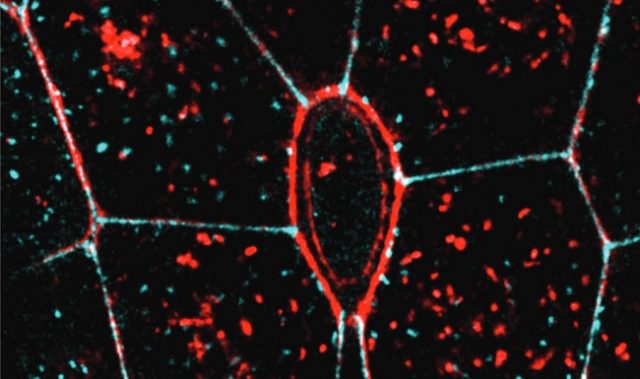
This technique allowed the researchers to observe organized patterns of motor protein filaments within large, cable-like protein structures known as stress fibers. Like ropes, these cables are made of many individual filaments, held together by various cross-linking proteins.
By watching the cytoskeleton form over time, the researchers observed how myosin II filaments arranged into stacks that ran perpendicular to the large parallel stress fibers. These stacks alternated with regions of the cross-linking protein a-actinin, which tethers individual filaments together to produce the protein cable.
How myosin II filaments come to be stacked together within the bundled stress fibers remains to be fully defined. However, one observation from this study that may hold the answer, is the long range movement of myosin II filaments towards each other. As the researchers propose, this attraction may result from contractile or elastic forces generated by the myosin filament stacks, which can transmit through the surrounding cytosol to individual filaments that are otherwise isolated.
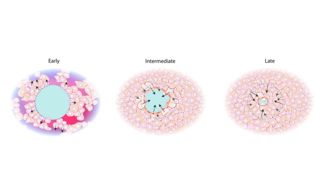
The stacking of myosin II filaments in non-muscle cells like the fibroblast is an intriguing element in the self-organization of the cytoskeleton and the overall architecture of the cell. Fibroblast function requires the cell to be able to stretch, generate cytoskeletal protrusions and move to other regions of the connective tissue. The assembly and organization of myosin II into stacks permits the fibroblast to fulfil these cellular processes.
The results show that the architecture of the cytoskeleton is specialized for force generation and sensing, even in non-muscle cells. In both cases contractile and elastic forces are integral in establishing a functional cytoskeleton, and once formed, a pattern of repeating protein-based contractile proteins becomes evident. However, unlike in muscle cells, these structures continuously assemble and disassemble in non-muscle cells, allowing them to adapt their function, shape, and direction of movement according to the environment they find themselves in.
As observed in this study, even non-muscle cells require strength to pull against their surroundings and fight their way through often sticky environments. This strength comes from a highly refined system of filaments and motor proteins. Although not as strong as those found in muscle cells, their organization in non-muscle cells allows them to remain responsive to changes in the environment, whilst providing just the right amount of force to carry out their functions.
The article can be found at: Hu et al. (2017) Long-range Self-Organization of Cytoskeletal Myosin II Filament Stacks.
———
Source: National University of Singapore; Photo: Shutterstock.
Disclaimer: This article does not necessarily reflect the views of AsianScientist or its staff.