AsianScientist (Jan. 26, 2017) – Researchers from the Mechanobiology Institute at the National University of Singapore have described how dying cells detach and are expelled from a tissue. Their findings, published in Development, could help biomedical engineers develop better artificial skin.
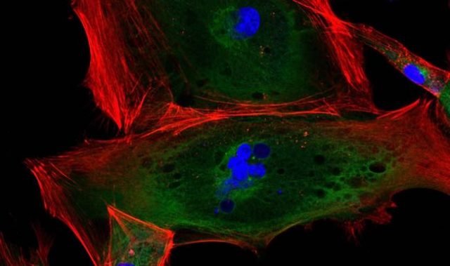
The integrity of any tissue is crucial to its function, and this is especially the case with epithelial tissue which lines the surface of our organs and body cavities. Just as a plastic bag would leak if it was punctured, so too would epithelial tissue if cells are damaged through injury or disease. Cells are also prone to dying when they are damaged and even the smallest holes left when a single cell dies must be filled immediately in order to maintain the integrity of the tissue.
However, the dying cell needs to be detached from its neighbors and expelled, in a process known as cell extrusion. If this does not occur, the dying cell is stacked in the tissue and causes further damage to the neighboring healthy tissue. Removing a dying cell from a tissue without compromising the tissue integrity is therefore not a trivial process.
To understand how dying cells are removed from epithelial tissue while the surrounding tissue remains intact and functional, researchers from the Mechanobiology Institute furthered their earlier investigations by viewing cells in a developing fruit fly pupae using live imaging.
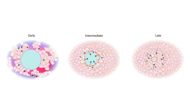
They found that proteins that connect the dying cell to its neighbors decrease, temporarily reducing the tension around the cell and allowing it to be released from the tissue. The hole left behind is then closed by a protein-based cable that passes through each cell surrounding the hole.
The contraction of the cable then rebuilds tissue tension, which forces the cells that surround the hole to become stretched. Stretching the cells brings them within proximity of the cells on the other side of the hole, with which they can form new cell-cell contacts. This eventually reseals the gap that transiently formed to let the dying cell go.
Ultimately, these findings provide new insights into the dynamic cellular processes that occur when tissue integrity is compromised. With tissue engineering reliant on our understanding of how our bodies naturally form and maintain healthy tissue, the findings from this study will help scientists better implement engineered tissues in the future.
The article can be found at: Teng et al. (2017) Remodeling of Adhesion and Modulation of Mechanical Tensile Forces During Apoptosis in Drosophila Epithelium.
———
Source: National University of Singapore.
Disclaimer: This article does not necessarily reflect the views of AsianScientist or its staff.