AsianScientist (Jun. 26, 2015) – Mapping the human brain’s network of interconnections—known as the connectome—is typically done with help from computational tools because recreating interconnections between different brain regions has been challenging in the lab.
Now, researchers at the Okinawa Institute of Science and Technology Graduate University (OIST) have developed a method to recreate connections between neurons from two different brain areas in a dish. Their findings have been published in Frontiers in Systems Neuroscience.
Researchers from OIST’s Brain Mechanism for Behavior Unit, Neurobiology Research Unit, as well as the Physics and Biology Unit collaborated on this study, using neurons from embryos of mice to study the first connections between different brain compartments develop at the embryonic stage.
Previously, the team had shown that culturing cortical and striatal cells within the same compartment resulted in an artificial connectomic system as these cortical and striatal neurons grew connections in all directions in a disorganized manner.
Inside a living brain, neurons within the cortex and striatum form dense interconnections with neurons within their respective brain compartments. There is only one-way electrophysiological traffic, from cortical cells to striatal cells via the formers’ axons. This one way street had been difficult to recreate in culture because striatal cells tend to die out when they do not receive electrophysiological impulses from cortical cells.
To overcome these challenges and yet keep the cortex and striatum cultures in separate compartments, the research team paired cortical and striatal compartments on the surface of multiple multi-electrode arrays (MEAs) and kept the cultures alive for three weeks.
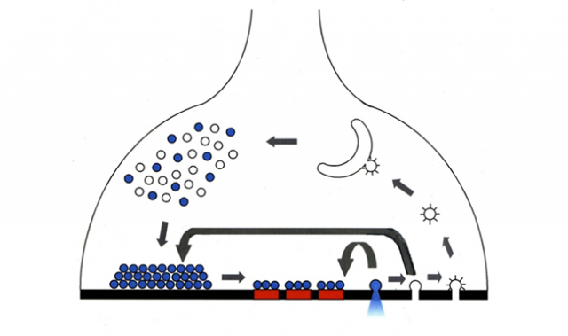
MEAs are tiny rectangular devices that consist of evenly spaced metal bumps arranged in a grid, all of which serve as electrodes. Each metal bump can transmit the measure of the electrical activity over it. Because many neurons talk to each other simultaneously, mathematical techniques are used to sort the signals from all active electrodes on a MEA to determine which groups are sending and receiving feedback from other groups.
After three weeks, enough axons had grown between the cortical and striatal compartments. The MEAs were then hooked up to a system to measure the bursts of electrical activity occurring at the different electrodes. While this system was recording, a cut was performed between the cortical and striatal compartments, severing the axons that had grown between them.
When this was done, electrical activity was snuffed out almost completely around the electrodes within the striatal compartment. The pattern of electrical activity between electrodes inside the cortical compartment was relatively unaffected, suggesting the interconnectivity between the cortical neurons was unchanged. This demonstrated that there were negligible upstream connections forming between striatal and cortical cells, and a working corticostriatal network had been recreated.
“We can also introduce a third compartment into this setup, possibly more. This would allow connections to grow between multiple types of neurons found in distant parts of the brain,” said Dr. Marianela Garcia-Munoz, group leader of the Brain Mechanism for Behavior Unit.
The article can be found at: Garcia-Munoz et al. (2015) Rebuilding A Realistic Corticostriatal “Social Network” From Dissociated Cells.
———
Source: Okinawa Institute of Science and Technology Graduate University.
Disclaimer: This article does not necessarily reflect the views of AsianScientist or its staff.