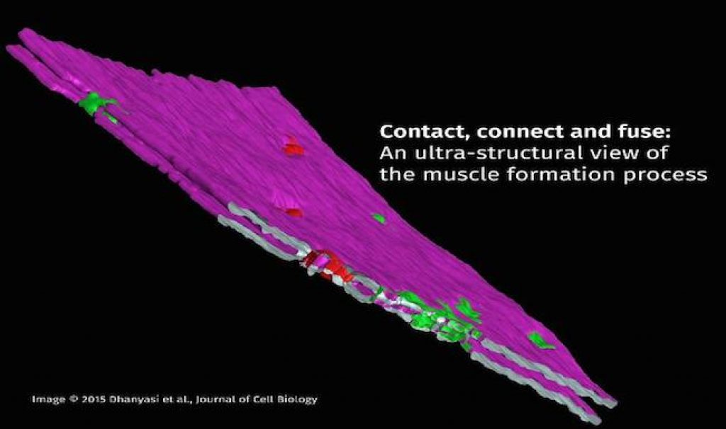
AsianScientist (Nov. 18, 2015) – Researchers have uncovered the way muscles develop and repair themselves in unprecedented detail, using electron microscopic techniques to show each distinct step in the fusion of myoblasts and muscle cells during the formation of flight muscles in the fruit fly (Drosophilia). Their results have been published in the Journal of Cell Biology.
When a person is running or working and tears their muscle, the injury causes dormant myosatellite cells to awaken and divide into myoblasts, which in turn fuse with muscle cells to repair damaged muscles. Previously, it was difficult to observe the fusion process between myoblasts and muscle cells.
To uncover more details of myoblast fusion, researchers at India’s National Centre for Biological Sciences (NCBS), Tata Institute of Fundamental Research (TIFR) collaborated with scientists at the Weizmann Institute of Science. To study the process at a high resolution, it was necessary to generate electron microscope (EM) images. This allowed researchers to reconstruct the steps in the dynamic process of myoblast fusion, where the structure of the fusing membranes was closely examined.
Most knowledge about the fusion of myoblasts to form multinucleate muscle fibers has been gleaned from studies on the development of tubular muscles of Drosophila larvae. However, the mechanisms governing myogenesis (formation of muscle fibers) in striated muscles of the skeletal musculature in vertebrates is not very clear.
The present study focuses on the events occurring in myoblast fusion in Drosophila during the formation of flight muscles. These muscles serve as a particularly attractive model, since their developmental program and muscle fiber organization resemble key aspects of vertebrate skeletal myogenesis.
The research built on previous investigations into the role of various fusion proteins using confocal microscopy in the myogenesis of flight muscles of Drosophila, said Dr. Nagaraju Dhanyasi, the first author of the paper. The earlier research had established the presence of stem cells in Drosophila muscles similar to the satellite cells in vertebrate muscles.
“This set the stage for addressing questions on vertebrate muscle regeneration in a Drosophila model system,” said Dhanyasi. “With the advent of RNAi technology, this problem was solved, and we were able to study the myoblast fusion process in great detail.”
The study shows that the fusion of myoblasts to existing muscle fibers, called myotubes, follows a set of distinct stages that requires communication between transmembrane elements and the actin cytoskeleton. An elegant series of experiments allowed the researchers to delineate the ultra-structural details of a series of discrete steps in this event.
The process begins with myoblasts binding to the surface of an existing myotube with the help of a host of cell adhesion proteins, a process known as apposition. Following apposition is a flattening of the myoblast membrane to increase its contact surface with the myotube. This step has been shown to require elements of the cell cytoskeletal machinery.
The third step in this process is a crucial one where the myoblast membrane and the myotube surface are brought very close to each other in a condition known as ‘tight apposition.’ The tight apposition forms multiple areas of cell-to-cell contacts called ‘nascent pore sites.’ These contact areas form fusion pores, where the cell membranes of the myoblast and myotubes merge. The fusion pores eventually expand until the myoblast is fully incorporated into the growing myotube.
Future studies are likely to involve detailed investigations on the mechanisms of fusion pore formation and in discovering the molecular players involved in pore formation.
“We believe that the cellular and molecular mechanisms uncovered in this study, and in future studies are highly conserved, and therefore also applicable in vertebrate systems. This study is therefore likely to provide key insights into understanding muscle development and repair processes”, said Dhanyasi.
The article can be found at: Dhanyasi et al. (2015) Surface Apposition and Multiple Cell Contacts Promote Myoblast Fusion in Drosophila Flight Muscles.
———
Source: Tata Institute of Fundamental Research.
Disclaimer: This article does not necessarily reflect the views of AsianScientist or its staff